
CBCT
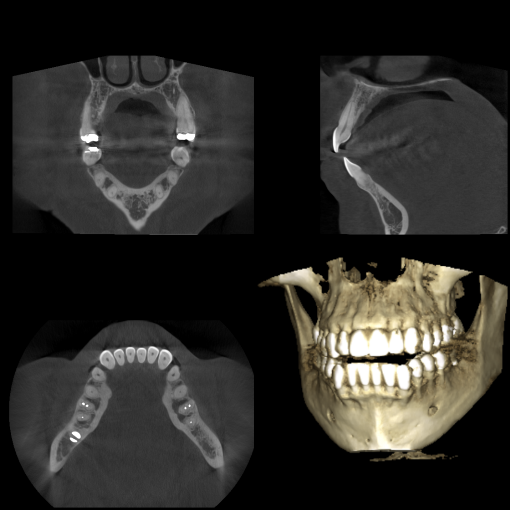
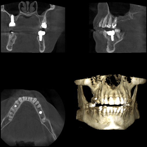
At our practice, we use Cone Beam Computed Tomography (CBCT). This very impressive piece of imaging machinery is capable of identifying many issues and structures that a normal x-ray is not. With traditional intraoral x-rays, the image is in 2D, allowing us to see only one viewpoint. With CBCT, we are able to see the entirety of the image in 3D, allowing us to better diagnose and treat our patient. To obtain the 3D x-ray, you will stand or sit in a chair with your chin on a small ledge. Once positioned in the machine, it will rotate around your entire head taking a full 360 degree view of the teeth, head, sinuses and bones.
The ability to view the full structure of your head as a whole is very informative to the dentist. It will allow us to see any potential problems and make sure that everything is functioning as it should be. CBCT is very useful in planning dental implant placement, completing difficult root canal therapy procedures, and many other procedures in dentistry. We are excited to bring this technology to our practice to better serve you!